
Two decades of kidney pathology are coming to Bigpicture
The Medical University of Vienna has started uploading kidney pathology data to Bigpicture and expects its contribution to eventually reach around 115,000–130,000 images. Covering a broad spectrum of kidney diseases, multiple stains and sequential tissue sections, the collection opens new possibilities for AI validation, benchmarking and renal pathology research.
Bigpicture is operational and already contains data from partners. Vienna’s contribution will add a substantial kidney pathology resource built from almost 20 years of routine diagnostic material. For Prof. Renate Kain, who oversees data contribution across WP3, the experience has reinforced one important point: “There is no path that fits all.” Contributors work with different archives, systems, expertise and legal and ethical frameworks. Bigpicture can provide common infrastructure and accumulated experience, but each institution still must connect its own environment to it.
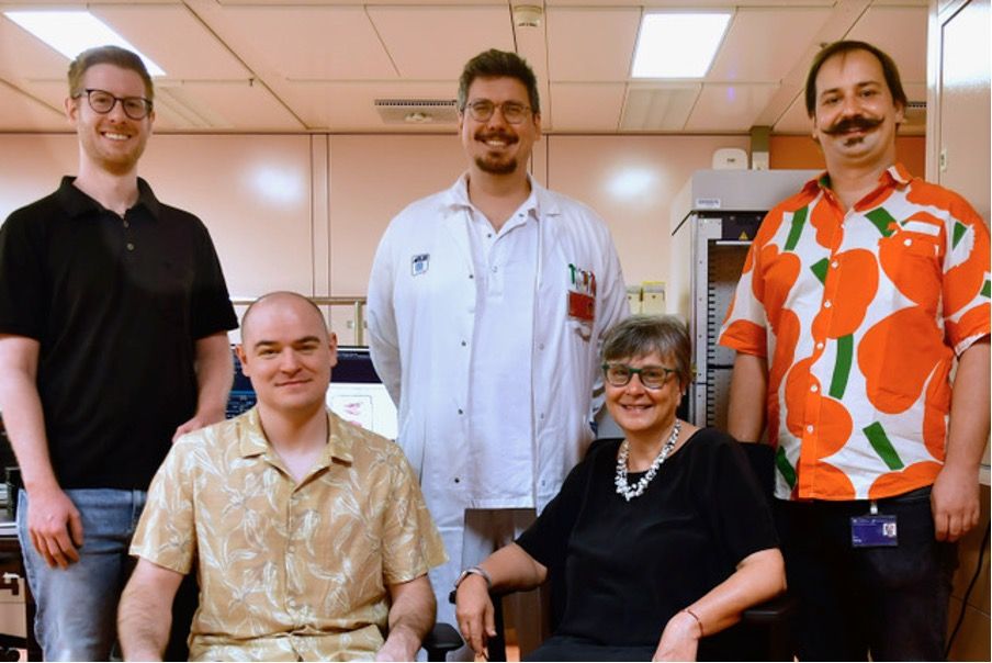
Prof. Renate Kain MD, PhD, Head of the Department of Pathology at the Medical University of Vienna, Bigpicture Work Package 3 co-lead and coordinator of the renal node.
Maximilian Köller MD, Bigpicture Work Package 3, and metadata taskforce member, pathologist, coordinating much of Vienna’s local dataset selection, quality control and Bigpicture contribution workflow.
Maximilian Gletthofer MSc, working on data science, software and the local tools used to collect, validate and prepare data for Bigpicture.
DI Christoph Stroblberger, part of the Digital and Computational Pathology group, working on software, data infrastructure and dataset creation.
Felix Küng PhD, mathematician supporting areas including case pre-selection and quality control.
The work also depends on Vienna’s wider kidney pathology team and the technical staff responsible for scanning the material.
What makes the Vienna collection interesting for research?
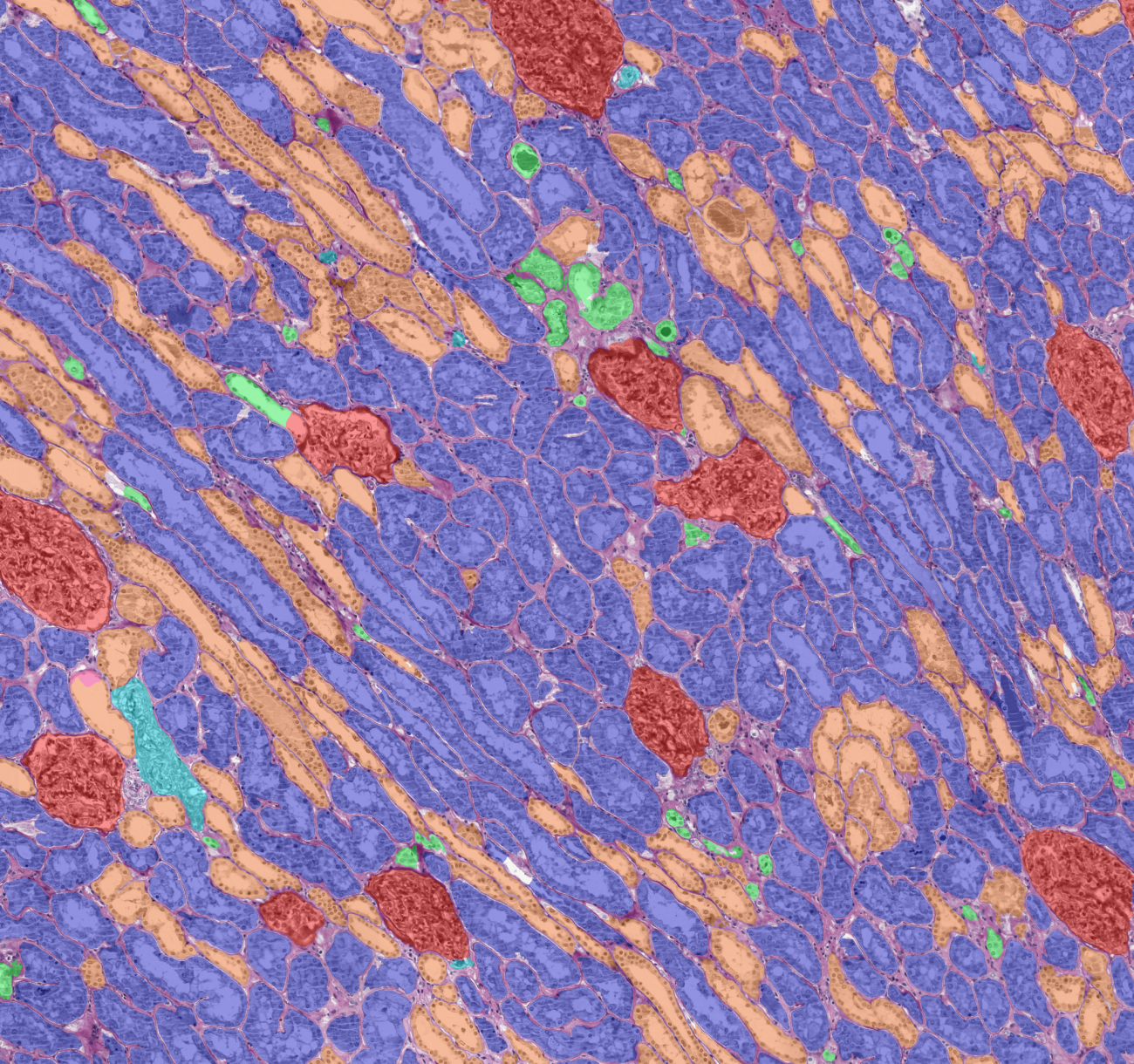
The Medical University of Vienna is retrospectively digitizing its kidney pathology archive rather than building a dataset around one narrowly defined research question. The collection therefore covers almost the full morphological and disease spectrum of non-neoplastic kidney pathology, including kidney transplantation cases, seen at the university hospital over roughly 20 years.
Maximilian (K) describes this as “data as it appears in the wild.” Instead of containing only clear, text book examples of a disease, the material includes the variation found in routine diagnostics, from morphology and staining to artefacts and changes over time. That makes it particularly relevant for testing how algorithms perform beyond highly streamlined research datasets.
Kidney pathology also provides unusually rich staining information. The archive systematically includes stains such as periodic acid-Schiff, trichrome, elastic van Gieson and silver, as well as immunohistochemical stains (e.g. IgG, IgM, IgA, C3, C1q, C4d among other more rarely used). Researchers can therefore investigate not only disease patterns but also information and variation across different stains.
A further feature is the way kidney biopsies are processed. Cases often contain more than 20 sequential tissue sections. Because these sections follow each other through the tissue, the team sees potential for future three-dimensional reconstruction and analysis of kidney biopsy cores.
The data is being organized into disease-focused datasets rather than uploaded simply in chronological batches. Researchers interested in IgA nephropathy, for example, can identify that collection separately from other forms of glomerulonephritis. Of special note is the dedicated strong focus on transplantation pathology at Vienna, with a large upcoming dedicated dataset including serial biopsies and over 30.000 images. Furthermore, datasets will continuously be expanded as further suitable data becomes available.
For future users: what this collection makes possible
- Validate and benchmark AI models on routine kidney pathology data
- Work with disease-specific datasets across a broad spectrum of kidney diseases, including transplantation cases
- Investigate different stains and staining variability
- Combine data from different centres to study inter-laboratory variation and model robustness
- Explore three-dimensional reconstruction using sequential tissue sections
- Develop dedicated collaborations in which additional clinical information can be connected to the pathology data where appropriate.
The base contribution is primarily pathology-focused. Additional clinical information is therefore not part of each dataset but could be added in specific research collaborations.
For future contributors: start with your own data
Vienna’s experience also highlights where much of the work in a large contribution actually sits: before the data reaches Bigpicture. Clinical information, research databases, scanned images and metadata often live in different local systems. The team at the Medical University of Vienna therefore developed tools to bring those sources together, check slide labels and metadata, and allow pathologists to review the information before deciding whether a case is suitable for a dataset. This is particularly important at scale. Scanning thousands of slides before establishing how they will be connected to the correct identifiers and metadata can create much more work later. For Maximilian (G), the approach is straightforward: “You really need to go step by step.”
Vienna’s experience points to four practical lessons:
- Bring the right expertise together. Large-scale contribution needs pathology, scanning and software/data expertise.
- Connect images and information from the start. Establish how slides, identifiers and metadata will be linked before scaling up.
- Begin with a small, robust workflow. Get the basics working reliably before adding more data sources and functionality.
- Design around the Bigpicture tools early. Contributors starting now can build their local process around the tools already available rather than developing separate solutions first.
This is also where Vienna’s experience shows how far the common infrastructure has come. Maximilian (K) describes the Bigpicture tools themselves as straightforward once local data can be connected to them: “They work perfectly well. It’s very easy to work with them.” The challenge is therefore less about finding one universal upload recipe and more about creating a reliable bridge between each contributor’s local environment and Bigpicture.
Part of Vienna’s collection is already being uploaded, with further disease-focused datasets to follow. For researchers, this adds something distinctive to Bigpicture: decades of kidney pathology with broad disease variation including transplantation cases, multiple stains and sequential sections, organised so that specific disease collections can be found and studied. It is a substantial addition to an already operational repository, and one that creates new possibilities for testing, comparing and developing computational approaches in kidney pathology.
Latest news

What does it take to turn 120,000 clinical pathology slides into data that researchers can actually use...
Read more
Being unique is one thing. Being valuable enough for organizations to rely on is another. Dr. Page...
Read more
The biggest hurdle for pathology AI may no longer be building the technology, but proving it works...
Read more